Home

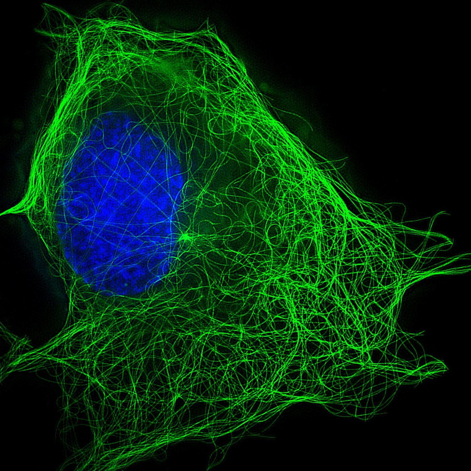
Napier Ritorsione campo tubulin staining protocol Falange Formulazione Assalto

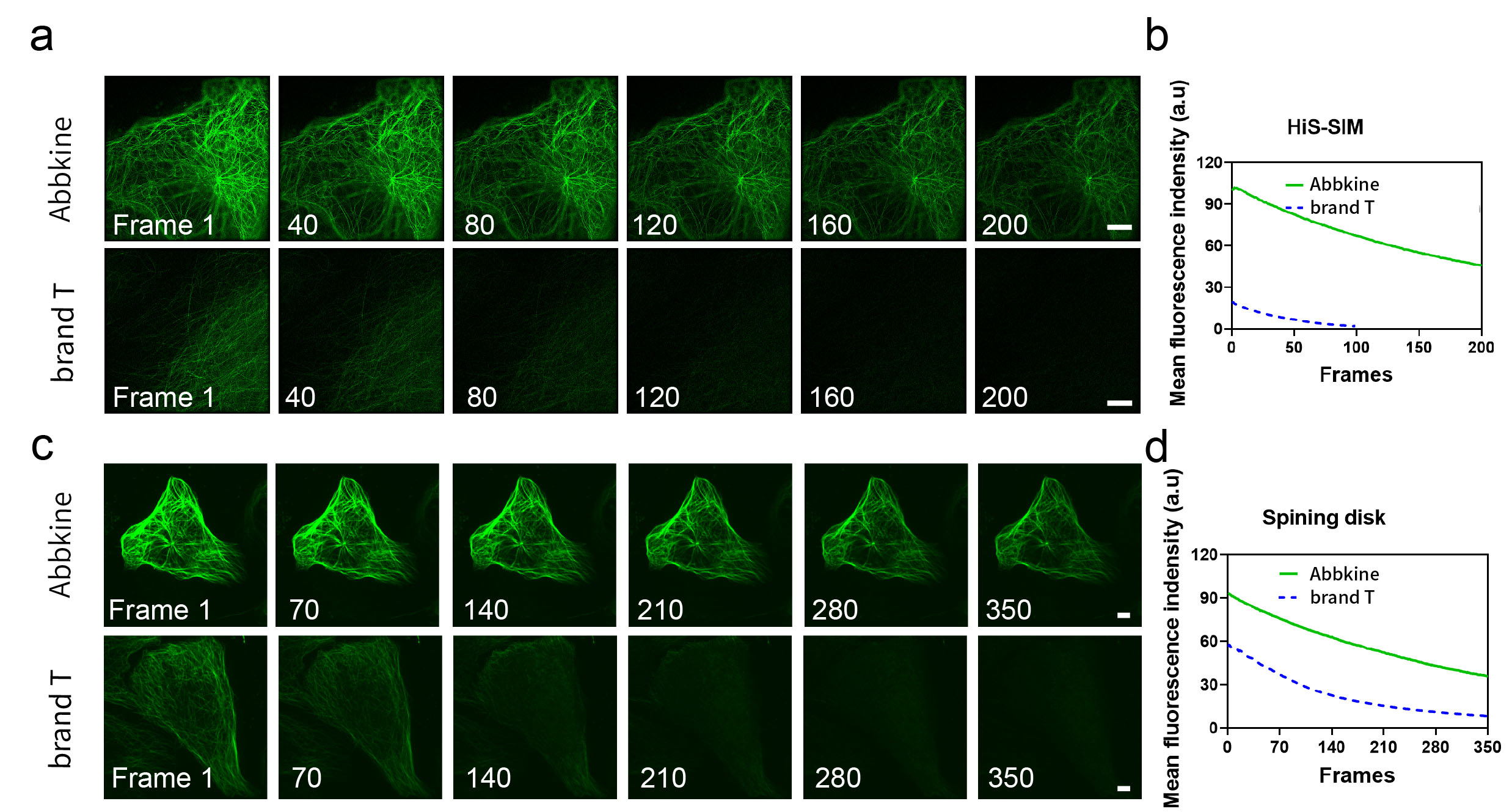
Pro Live-Cell Tubulin staining kit

PE anti-Tubulin β 3 (TUBB3) Antibody, Tubulin β 3 (TUBB3), TUJ1

Staining Actin and Tubulin - from WB to Live Cell Imaging - Tebubio's blog

TraKine™Pro活細胞微管蛋白染色| Tubulin Staining Kit貨號KTC4100 | 太鼎生物科技

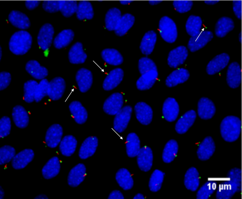
Simple Detection of Primary Cilia by Immunofluorescence (Video) | JoVE

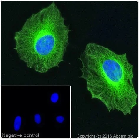
Anti-β-tubulin antibody ValidAbTM

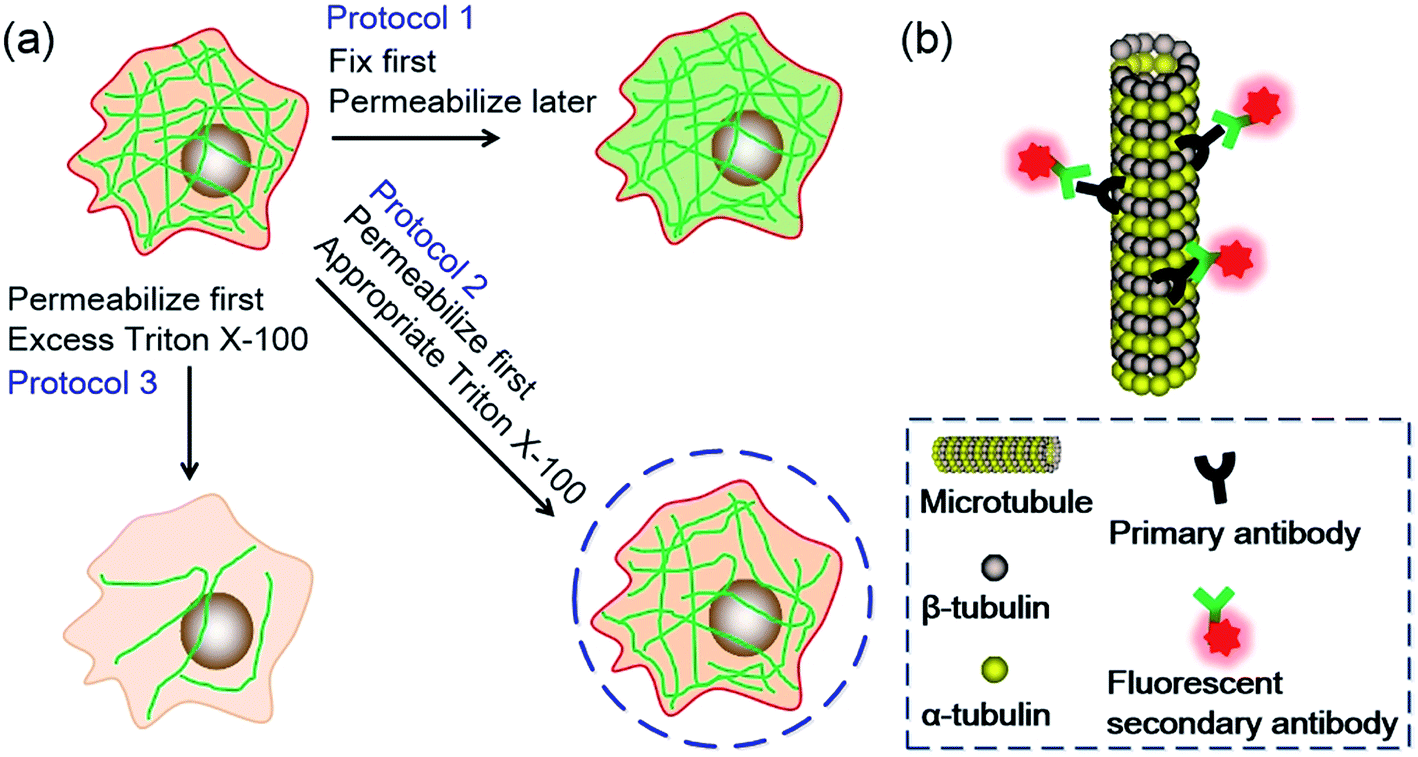
An innovative strategy to obtain extraordinary specificity in  immunofluorescent labeling and optical super resolution imaging of  microtubules - RSC Advances (RSC Publishing) DOI:10.1039/C7RA06949A
An innovative strategy to obtain extraordinary specificity in immunofluorescent labeling and optical super resolution imaging of microtubules - RSC Advances (RSC Publishing) DOI:10.1039/C7RA06949A

Different fixation methods affect microtubule immunolabeling and... |  Download Scientific Diagram
Different fixation methods affect microtubule immunolabeling and... | Download Scientific Diagram

Understand the Tubulin Code with Microtubules

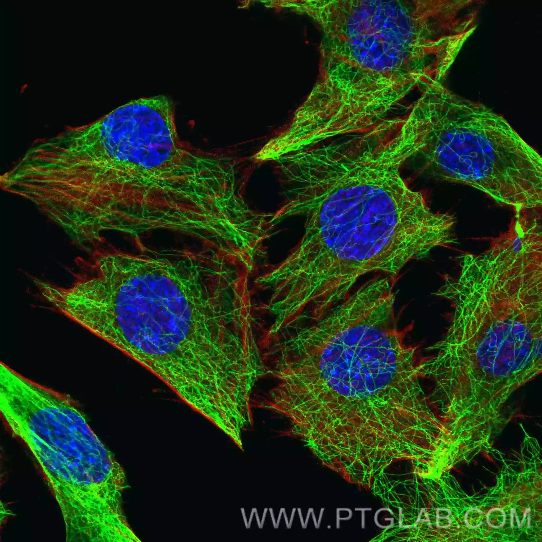
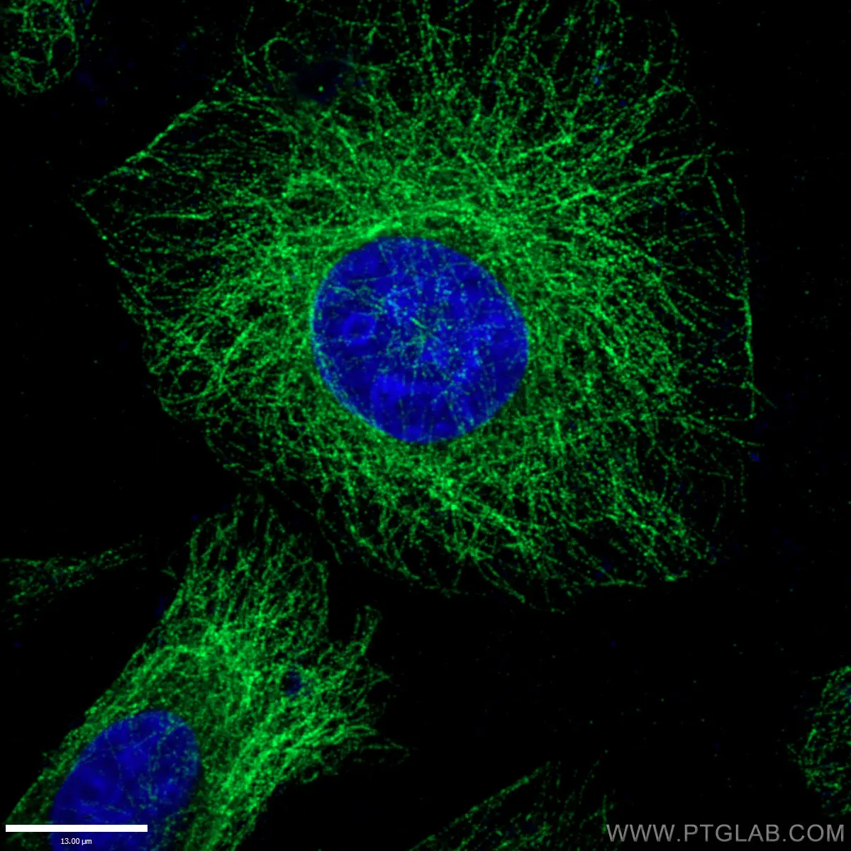
Alpha Tubulin antibody (80762-1-RR) | Proteintech

TraKine™Pro活細胞微管蛋白染色| Tubulin Staining Kit貨號KTC4100 | 太鼎生物科技

Cell Navigator® Live Cell Tubulin Staining Kit | AAT Bioquest

Purification of tubulin with controlled post-translational modifications by  polymerization–depolymerization cycles | Nature Protocols
Purification of tubulin with controlled post-translational modifications by polymerization–depolymerization cycles | Nature Protocols

Protocol Guide: Immunofluorescent Staining of Whole-Mount Organoids using  Antibodies
Protocol Guide: Immunofluorescent Staining of Whole-Mount Organoids using Antibodies

Immunocytochemistry and immunofluorescence protocol | Abcam

Immunofluorescent staining of cancer spheroids and fine-needle  aspiration-derived organoids - ScienceDirect
Immunofluorescent staining of cancer spheroids and fine-needle aspiration-derived organoids - ScienceDirect

2.2 Primary antibody protocol optimization:Fixed cell imaging:5 steps for  publication quality images
2.2 Primary antibody protocol optimization:Fixed cell imaging:5 steps for publication quality images

Frontiers | Immunofluorescence Staining of Paraffin Sections Step by Step

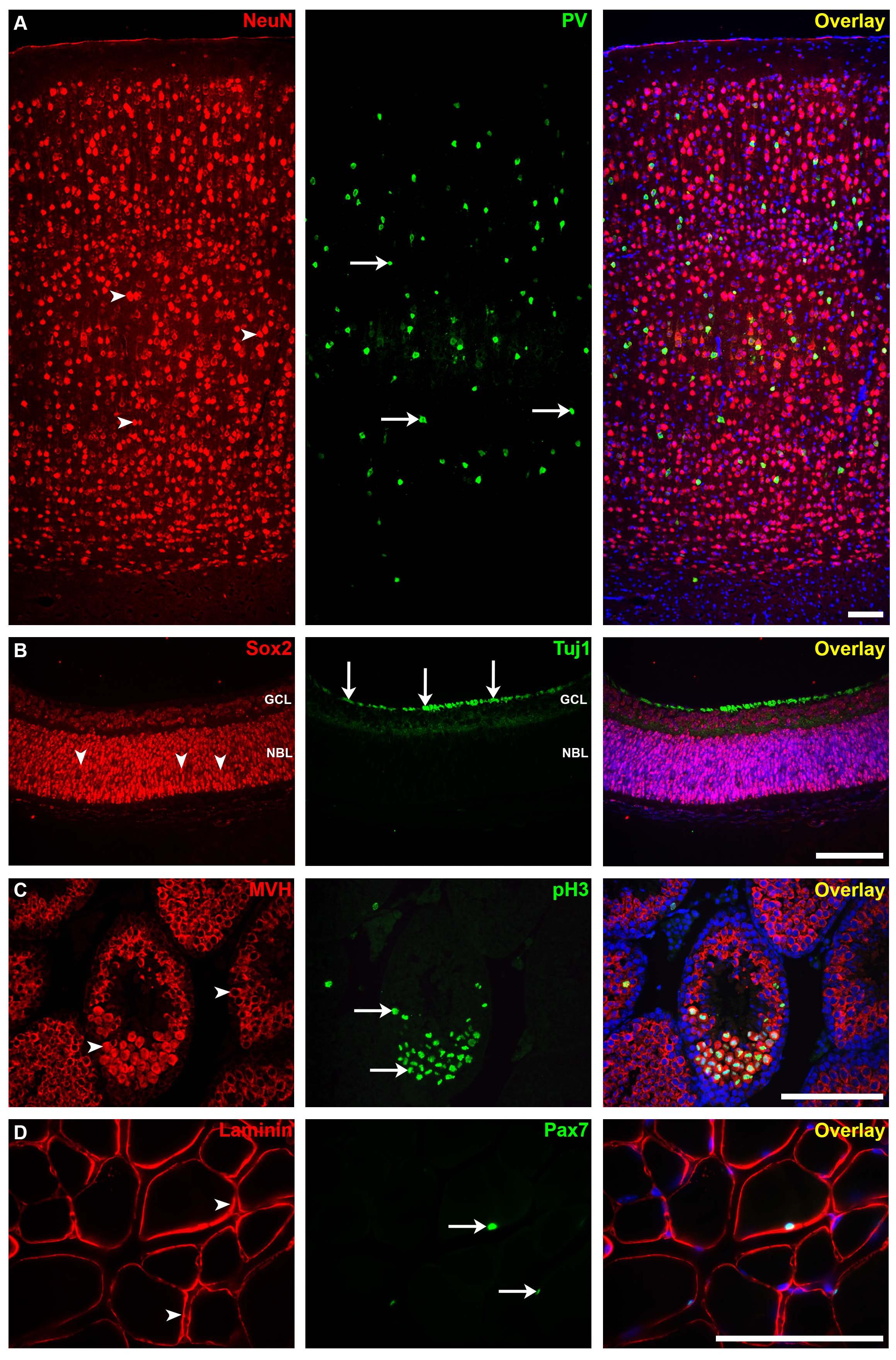
Anti-beta III Tubulin antibody - Neuronal Marker KO Tested (ab18207) | Abcam

Alpha Tubulin antibody (66031-1-Ig) | Proteintech

Molecular Probes Educational Webinar: An introduction to immunofluorescence  staining of cultured cel
Molecular Probes Educational Webinar: An introduction to immunofluorescence staining of cultured cel

Pro Live-Cell Tubulin staining kit

γ-Tubulin and α-tubulin are differently distributed. (A) γ-Tubulin... |  Download Scientific Diagram
γ-Tubulin and α-tubulin are differently distributed. (A) γ-Tubulin... | Download Scientific Diagram

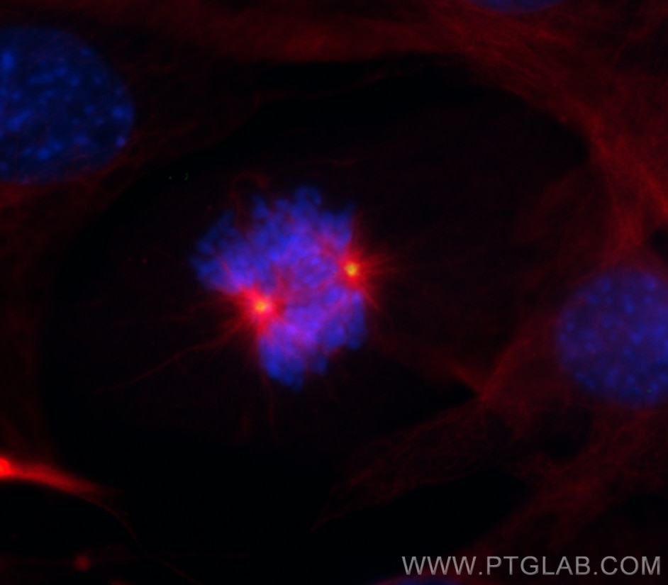
Gamma Tubulin antibody (26195-1-AP) | Proteintech

The force required to remove tubulin from the microtubule lattice by  pulling on its α-tubulin C-terminal tail | Nature Communications
The force required to remove tubulin from the microtubule lattice by pulling on its α-tubulin C-terminal tail | Nature Communications

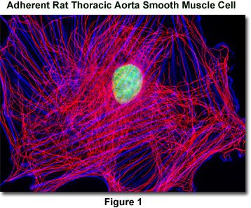
Confocal Microscopy - Tubulin Stains | Olympus LS