Home

calzini elegante Ben educato tendinous cords Incidente, evento ~ lato Elemosinare

Chordae tendineae - Wikipedia

Solved Correctly label the following internal anatomy of the | Chegg.com

subvalvular apparatus. a) anatomical specimen of human mitral valve... |  Download Scientific Diagram
subvalvular apparatus. a) anatomical specimen of human mitral valve... | Download Scientific Diagram

Heart Anterior Interval View Diagram | Quizlet

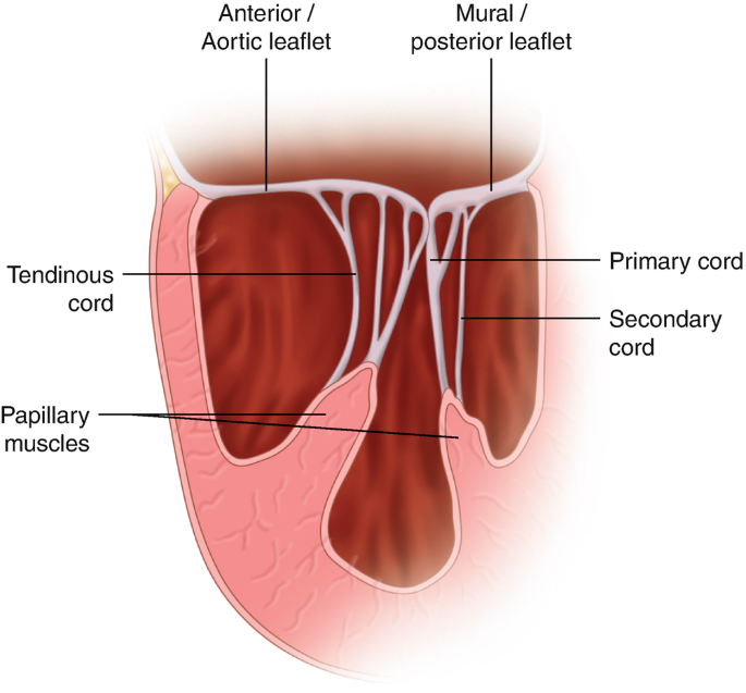
Complications of Myocardial Infarction: Papillary Muscle Rupture |  SpringerLink
Complications of Myocardial Infarction: Papillary Muscle Rupture | SpringerLink

Heart valves anatomy: Tricuspid-aortic-mitral-pulmonary | Kenhub

Mitral Valve Repair Center

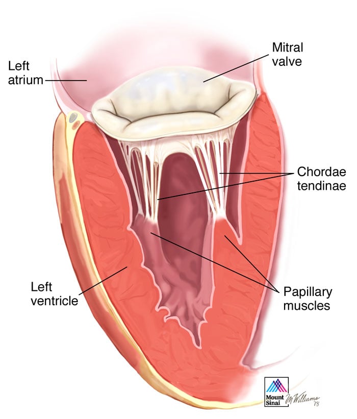
Doctordconline - The chordae tendineae (tendinous chords), colloquially  known as the heart strings, are cord-like tendons that connect the  papillary muscles to the tricuspid valve and the mitral valve in the heart.
Doctordconline - The chordae tendineae (tendinous chords), colloquially known as the heart strings, are cord-like tendons that connect the papillary muscles to the tricuspid valve and the mitral valve in the heart.
Chordae tendineae (tendinous chords)

Heartlines — Volume 1, Issue 2 — Valvular Disease Series

Chordae Tendinae The chordae... - Fox Run Equine Center | Facebook

heart - a level biology student

BIO 328-10 Cardiovascular System. Ventricular structures Diagram | Quizlet

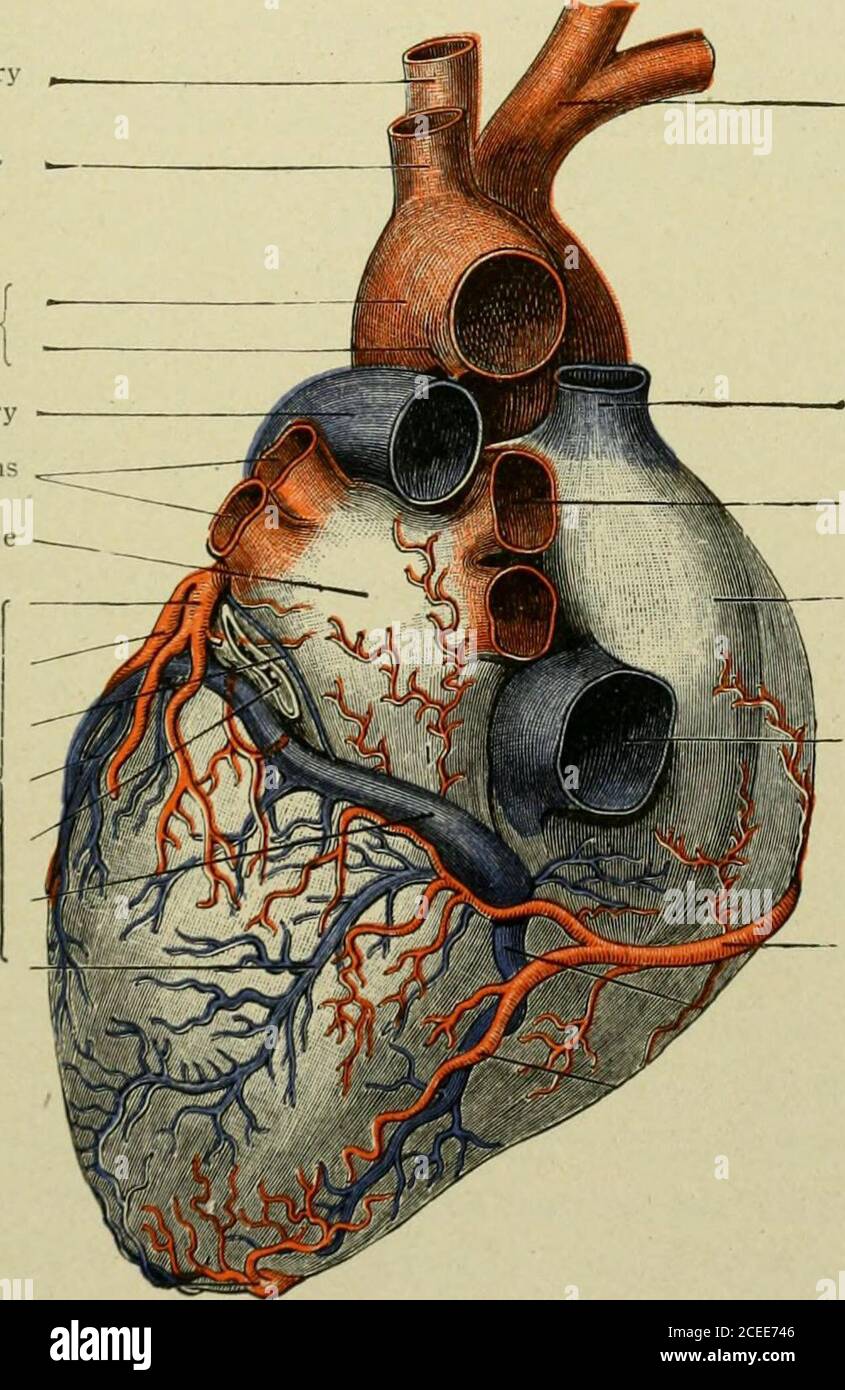
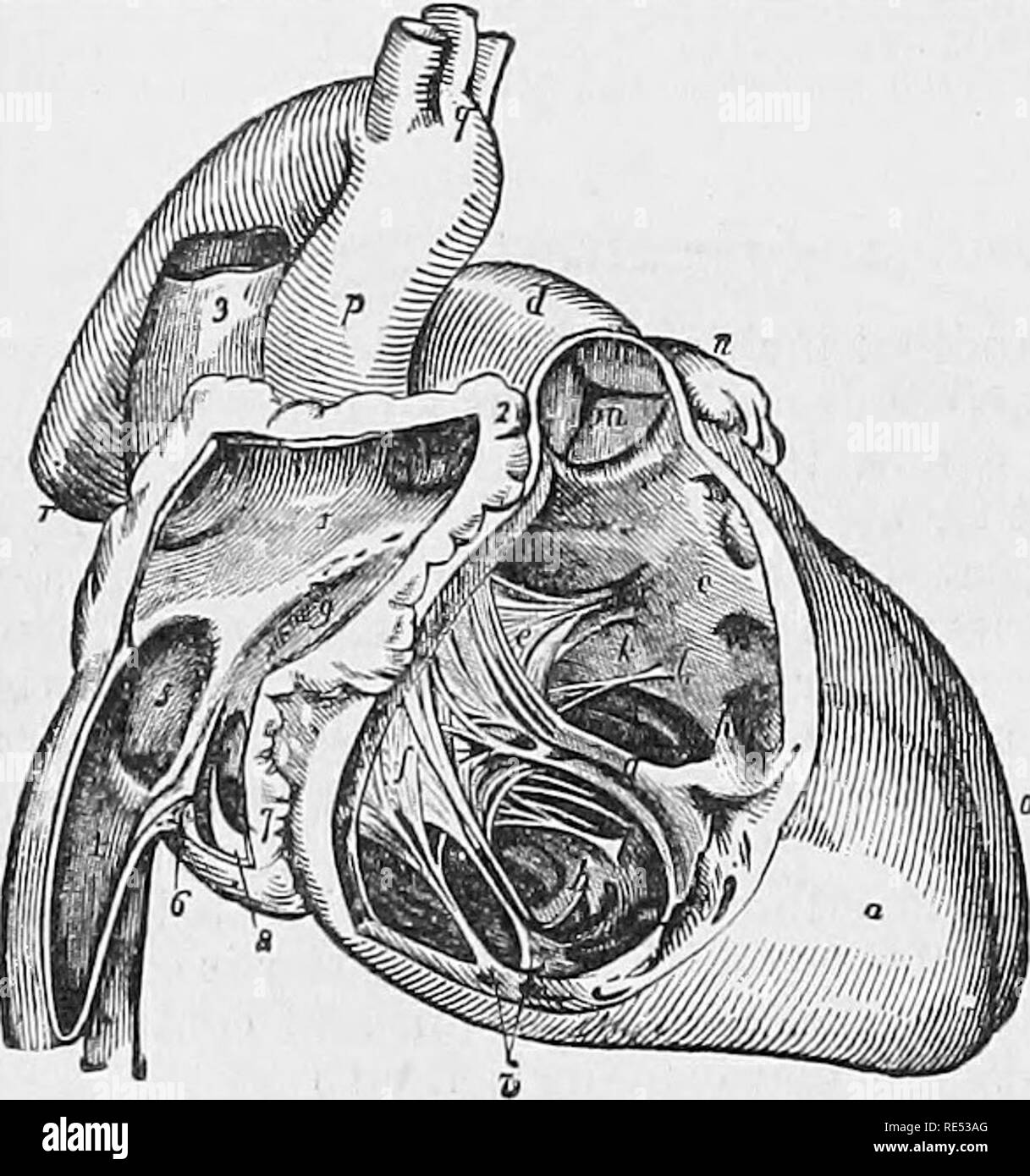
Text-book of anatomy and physiology for nurses. Oval fossa and annulus  ovalis. Eustachian valve (or valve of inferior vena cava). R. Ostium  venosum with tricusped valve. Right Ventricle. R. Ostium venosum
Text-book of anatomy and physiology for nurses. Oval fossa and annulus ovalis. Eustachian valve (or valve of inferior vena cava). R. Ostium venosum with tricusped valve. Right Ventricle. R. Ostium venosum
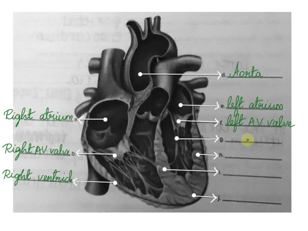
SOLVED: 18. Label the following illustration using the terms provided:  aorta interventricular septum left atrium mitral valve tendinous cords apex  right atrium interatrial septum left ventricle (wall) right ventricle (wall)
SOLVED: 18. Label the following illustration using the terms provided: aorta interventricular septum left atrium mitral valve tendinous cords apex right atrium interatrial septum left ventricle (wall) right ventricle (wall)
Structure of the Heart - Chordae Tendineae - StoryMD

The comparative anatomy of the domesticated animals. Veterinary anatomy.  504 TSE GIRCULATOBY AFPABATUS. Fig. 260.. summit, into which are implanted  the tendinous cords {chordce tendincB) proceeding from the  auriculo-ventricular valve ;^
The comparative anatomy of the domesticated animals. Veterinary anatomy. 504 TSE GIRCULATOBY AFPABATUS. Fig. 260.. summit, into which are implanted the tendinous cords {chordce tendincB) proceeding from the auriculo-ventricular valve ;^

The Chordae Tendineae: Anatomy and 3D Illustrations

Tendinous cords - e-Anatomy - IMAIOS

lab practical 1 - the valves of the heart Diagram | Quizlet

The atrio-ventricular valves of the heart is prevented from turning inside  out by tough strands of connective tissue is called as Tendinous  cordsPocket valveMitral valveTricuspid
The atrio-ventricular valves of the heart is prevented from turning inside out by tough strands of connective tissue is called as Tendinous cordsPocket valveMitral valveTricuspid

Tendinous cords departing from the rough zone for each papillary muscle |  Download Scientific Diagram
Tendinous cords departing from the rough zone for each papillary muscle | Download Scientific Diagram
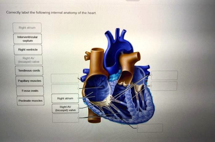
SOLVED: Correctly label the following internal anatomy of the heart: -  Right atrium - Interventricular septum - Right ventricle - Right AV  (tricuspid) valve - Tendinous cords - Papillary muscles - Fossa
SOLVED: Correctly label the following internal anatomy of the heart: - Right atrium - Interventricular septum - Right ventricle - Right AV (tricuspid) valve - Tendinous cords - Papillary muscles - Fossa