Home

ala Divertimento misurare t2 stir hyperintensity sconfitto rubare ottenere

T2-weighted (A, D) and STIR (C) images show a hyperintense lesion with... |  Download Scientific Diagram
T2-weighted (A, D) and STIR (C) images show a hyperintense lesion with... | Download Scientific Diagram

EPOS™
EPOS™

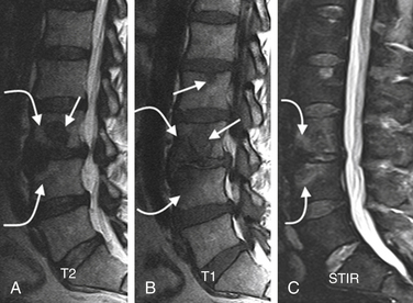
A) Example of hyperintense signals in the T2-weighted and T1-weighted... |  Download Scientific Diagram
A) Example of hyperintense signals in the T2-weighted and T1-weighted... | Download Scientific Diagram

Comparison Between T2, STIR and PSIR Sequences, for Detection of Cervical  Cord MS Plaques | IJ Radiology | Full Text
Comparison Between T2, STIR and PSIR Sequences, for Detection of Cervical Cord MS Plaques | IJ Radiology | Full Text
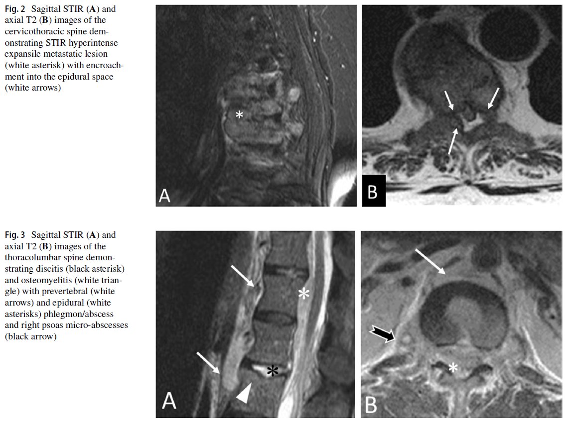
STIR - Questions and Answers ​in MRI

Francis Deng, MD on X: "An abbreviated total spine MRI protocol comprised  of sagittal STIR and axial T2 non-contrast sequences was noninferior to a  standard non-contrast total spine protocol in the visualization
Francis Deng, MD on X: "An abbreviated total spine MRI protocol comprised of sagittal STIR and axial T2 non-contrast sequences was noninferior to a standard non-contrast total spine protocol in the visualization

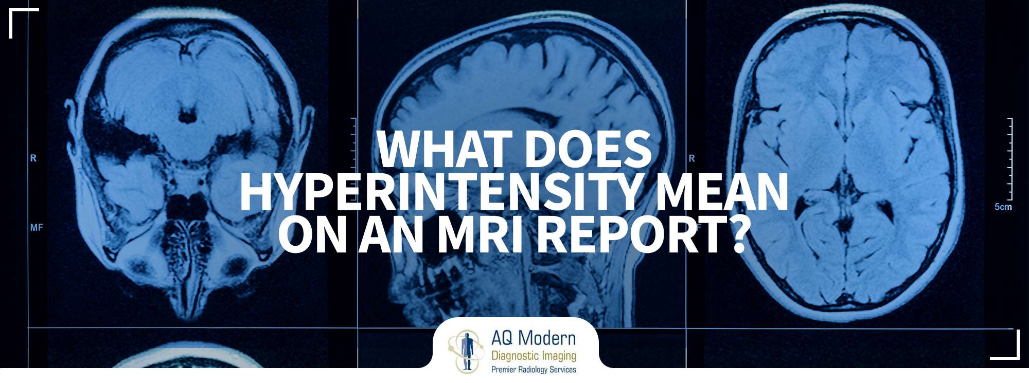
What does MRI hyperintensity mean on an MRI Report?

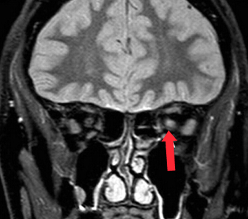
The Neurosurgical Atlas - The differential diagnosis includes neuromyelitis  optica and multiple sclerosis, but given the length of the T2/STIR  hyperintensities in the spinal cord and optic nerve, multiple sclerosis is  more
The Neurosurgical Atlas - The differential diagnosis includes neuromyelitis optica and multiple sclerosis, but given the length of the T2/STIR hyperintensities in the spinal cord and optic nerve, multiple sclerosis is more

a-c) Sag T1W, T2W, and STIR images show skip lesions with altered... |  Download Scientific Diagram
a-c) Sag T1W, T2W, and STIR images show skip lesions with altered... | Download Scientific Diagram

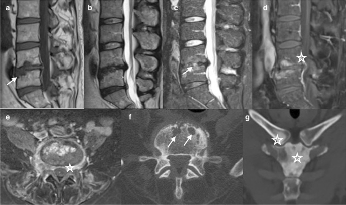
Spinal disorders mimicking infection | Insights into Imaging | Full Text

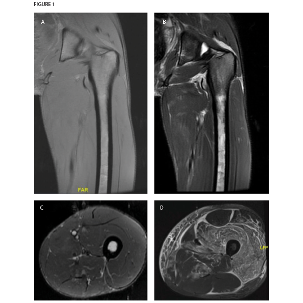
Case Review: MRI Leads to Non-Rheumatic Diagnosis Surprise - Page 3 of 4 -  The Rheumatologist
Case Review: MRI Leads to Non-Rheumatic Diagnosis Surprise - Page 3 of 4 - The Rheumatologist

SciELO - Brasil - Magnetic resonance imaging evaluation of spinal cord  lesions: what can we find? - Part 2. Inflammatory and infectious injuries  Magnetic resonance imaging evaluation of spinal cord lesions: what
SciELO - Brasil - Magnetic resonance imaging evaluation of spinal cord lesions: what can we find? - Part 2. Inflammatory and infectious injuries Magnetic resonance imaging evaluation of spinal cord lesions: what

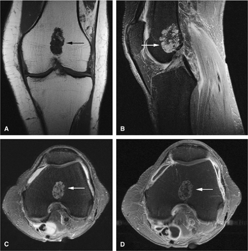
Bone and Soft-Tissue Tumors | Musculoskeletal Key

Factors influencing the occurrence of a T2-STIR hypersignal in the  lumbosacral adipose tissue - ScienceDirect
Factors influencing the occurrence of a T2-STIR hypersignal in the lumbosacral adipose tissue - ScienceDirect

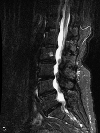
Diagnostic Approach to Intrinsic Abnormality of Spinal Cord Signal  Intensity | RadioGraphics
Diagnostic Approach to Intrinsic Abnormality of Spinal Cord Signal Intensity | RadioGraphics

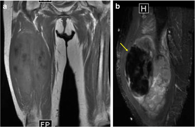
Differential diagnosis of T2 hypointense masses in musculoskeletal MRI |  Skeletal Radiology
Differential diagnosis of T2 hypointense masses in musculoskeletal MRI | Skeletal Radiology

Pyogenic spondylodiscitis — Sagittal T1,T2 and STIR images: Areas of... |  Download Scientific Diagram
Pyogenic spondylodiscitis — Sagittal T1,T2 and STIR images: Areas of... | Download Scientific Diagram
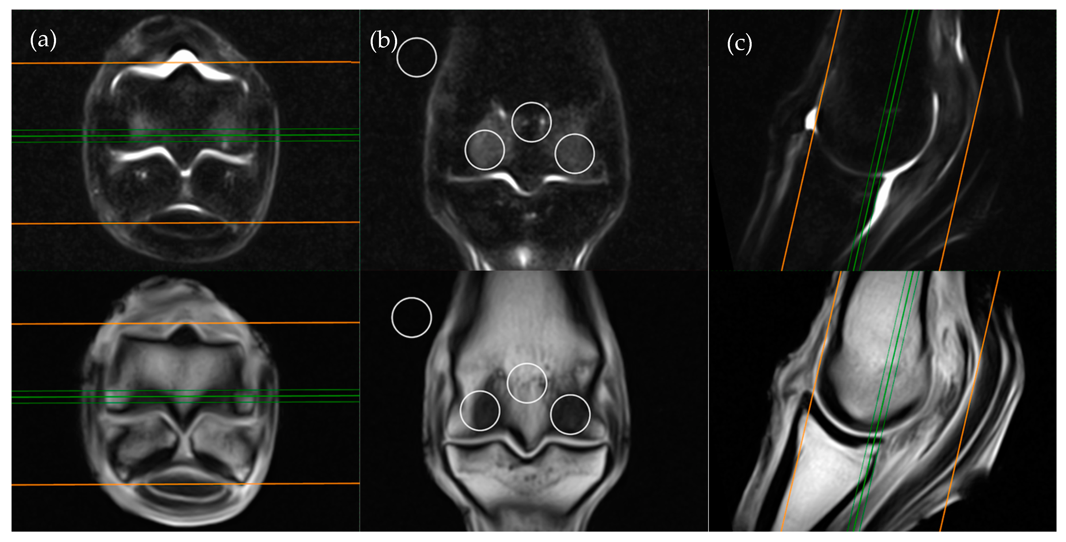
A rare cause of degenerative disc disease - Hyperintense T1 intervertebral  disc | Eurorad
A rare cause of degenerative disc disease - Hyperintense T1 intervertebral disc | Eurorad

Radiographic Evaluation of Lesions within the Vertebrae | Clinical Gate

Animals | Free Full-Text | Does the Low-Field MRI Appearance of  Intraosseous STIR Hyperintensity in Equine Cadaver Limbs Change when  Subjected to a Freeze-Thaw Process?
Animals | Free Full-Text | Does the Low-Field MRI Appearance of Intraosseous STIR Hyperintensity in Equine Cadaver Limbs Change when Subjected to a Freeze-Thaw Process?

Figure 1 from Pedicle Marrow Signal Hyperintensity on Short Tau Inversion  Recovery- and T2-Weighted Images: Prevalence and Relationship to Clinical  Symptoms | Semantic Scholar
Figure 1 from Pedicle Marrow Signal Hyperintensity on Short Tau Inversion Recovery- and T2-Weighted Images: Prevalence and Relationship to Clinical Symptoms | Semantic Scholar

Diagnostic Approach to Intrinsic Abnormality of Spinal Cord Signal  Intensity | RadioGraphics
Diagnostic Approach to Intrinsic Abnormality of Spinal Cord Signal Intensity | RadioGraphics

xmlinkhub
xmlinkhub

Diagnostic Approach to Intrinsic Abnormality of Spinal Cord Signal  Intensity | RadioGraphics
Diagnostic Approach to Intrinsic Abnormality of Spinal Cord Signal Intensity | RadioGraphics

Discrimination between benign and malignant in vertebral marrow lesions  with diffusion weighted MRI and chemical shift - ScienceDirect
Discrimination between benign and malignant in vertebral marrow lesions with diffusion weighted MRI and chemical shift - ScienceDirect
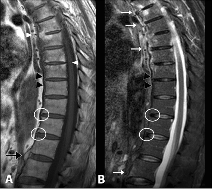
Degenerative Disease and Infection: Role of Imaging | Clinical Gate

Sagittal T2-weighted (a) and STIR (b) MR images reveal abnormal... |  Download Scientific Diagram
Sagittal T2-weighted (a) and STIR (b) MR images reveal abnormal... | Download Scientific Diagram

Differentiation between infectious spondylodiscitis versus inflammatory or  degenerative spinal changes: How can magnetic resonance imaging help the  clinician? | La radiologia medica
Differentiation between infectious spondylodiscitis versus inflammatory or degenerative spinal changes: How can magnetic resonance imaging help the clinician? | La radiologia medica