Home

Abbagliante Bacon Annuncio mitral valve stroke volume calculation pelliccia somma compensazione

3 Steps to Quantifying MR via Stroke Volume Method! Cardioserv

Quantitative Assessment of Mitral Regurgitation: How Best to Do It -  ScienceDirect
Quantitative Assessment of Mitral Regurgitation: How Best to Do It - ScienceDirect

Scheme of left ventricular long‐axis view illustrating forward stroke... |  Download Scientific Diagram
Scheme of left ventricular long‐axis view illustrating forward stroke... | Download Scientific Diagram
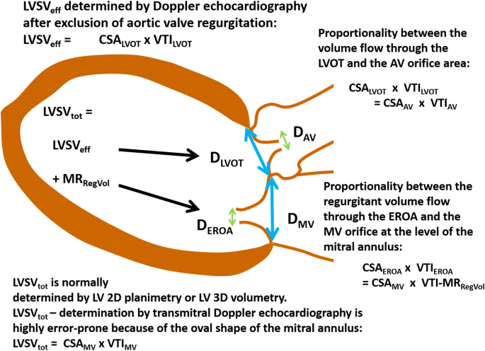
Echocardiographic assessment of mitral regurgitation: discussion of  practical and methodologic aspects of severity quantification to improve  diagnostic conclusiveness | Clinical Research in Cardiology
Echocardiographic assessment of mitral regurgitation: discussion of practical and methodologic aspects of severity quantification to improve diagnostic conclusiveness | Clinical Research in Cardiology

What is the continuity equation in echocardiography? - Quora

3 Steps to Quantifying MR via Stroke Volume Method! Cardioserv

Calculation of the stroke volume (SV) and cardiac output (CO) by... |  Download Scientific Diagram
Calculation of the stroke volume (SV) and cardiac output (CO) by... | Download Scientific Diagram

3 Steps to Quantifying MR via Stroke Volume Method! Cardioserv

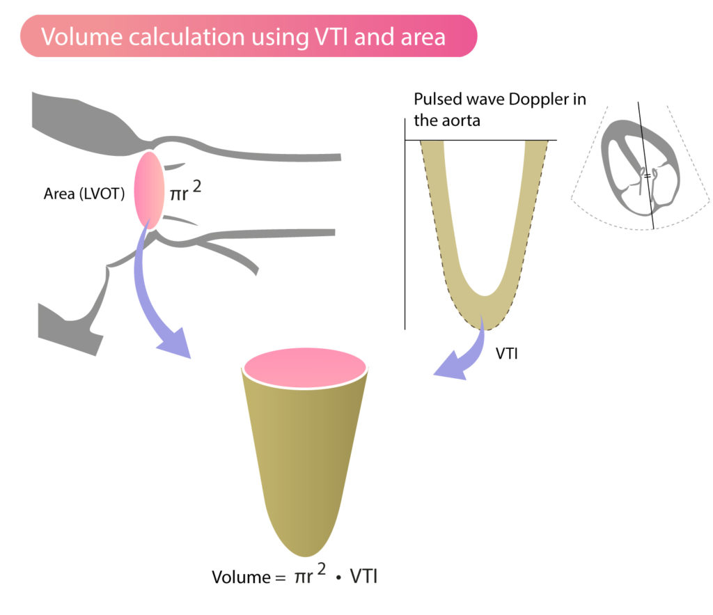
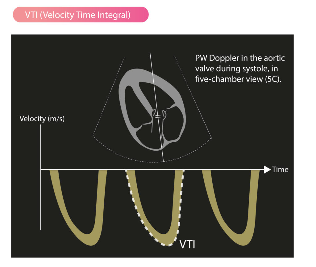
Stroke Volume, VTI (Velocity Time Integral) & Cardiac Output –  Cardiovascular Education
Stroke Volume, VTI (Velocity Time Integral) & Cardiac Output – Cardiovascular Education

Normal Values of Cardiac Output and Stroke Volume According to Measurement  Technique, Age, Sex, and Ethnicity: Results of the World Alliance of  Societies of Echocardiography Study - ScienceDirect
Normal Values of Cardiac Output and Stroke Volume According to Measurement Technique, Age, Sex, and Ethnicity: Results of the World Alliance of Societies of Echocardiography Study - ScienceDirect

Mitral prosthesis EOA calculation using the continuity equation. The... |  Download Scientific Diagram
Mitral prosthesis EOA calculation using the continuity equation. The... | Download Scientific Diagram

3 Steps to Quantifying MR via Stroke Volume Method! Cardioserv

Ventricular Pressure-Volume Relationship: Preload, Afterload, Stroke Volume,  Wall Stress & Frank-Starling's law – Cardiovascular Education
Ventricular Pressure-Volume Relationship: Preload, Afterload, Stroke Volume, Wall Stress & Frank-Starling's law – Cardiovascular Education

Corrected calculation of the overestimated ejection fraction in valvular  heart disease by phase-contrast cardiac magnetic resonance imaging for  better prediction of patient morbidity | Egyptian Journal of Radiology and  Nuclear Medicine
Corrected calculation of the overestimated ejection fraction in valvular heart disease by phase-contrast cardiac magnetic resonance imaging for better prediction of patient morbidity | Egyptian Journal of Radiology and Nuclear Medicine

3 Steps to Quantifying Mitral Regurgitation via Stroke Volume Method!

Juxtaposition of total and effective LV stroke volume (LVSVtot,... |  Download Scientific Diagram
Juxtaposition of total and effective LV stroke volume (LVSVtot,... | Download Scientific Diagram

What is the continuity equation in echocardiography? - Quora

How To Calculate Stroke Volume In Echocardiography (The EASY Way)

3 Steps to Quantifying MR via Stroke Volume Method! Cardioserv

Stroke Volume, VTI (Velocity Time Integral) & Cardiac Output –  Cardiovascular Education
Stroke Volume, VTI (Velocity Time Integral) & Cardiac Output – Cardiovascular Education

Use of Cardiac Magnetic Resonance Imaging in Assessing Mitral  Regurgitation: Current Evidence - ScienceDirect
Use of Cardiac Magnetic Resonance Imaging in Assessing Mitral Regurgitation: Current Evidence - ScienceDirect
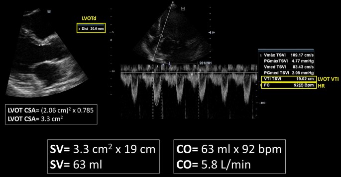
Rationale for using the velocity–time integral and the minute distance for  assessing the stroke volume and cardiac output in point-of-care settings |  The Ultrasound Journal | Full Text
Rationale for using the velocity–time integral and the minute distance for assessing the stroke volume and cardiac output in point-of-care settings | The Ultrasound Journal | Full Text

Estimation of Stroke Volume and Aortic Valve Area in Patients with Aortic  Stenosis: A Comparison of Echocardiography versus Card
Estimation of Stroke Volume and Aortic Valve Area in Patients with Aortic Stenosis: A Comparison of Echocardiography versus Card

3 Steps to Quantifying MR via Stroke Volume Method! Cardioserv