Home

Onesto Forma della nave Zoo di notte ihc nuclear staining Razionale Per dare il permesso di spessore

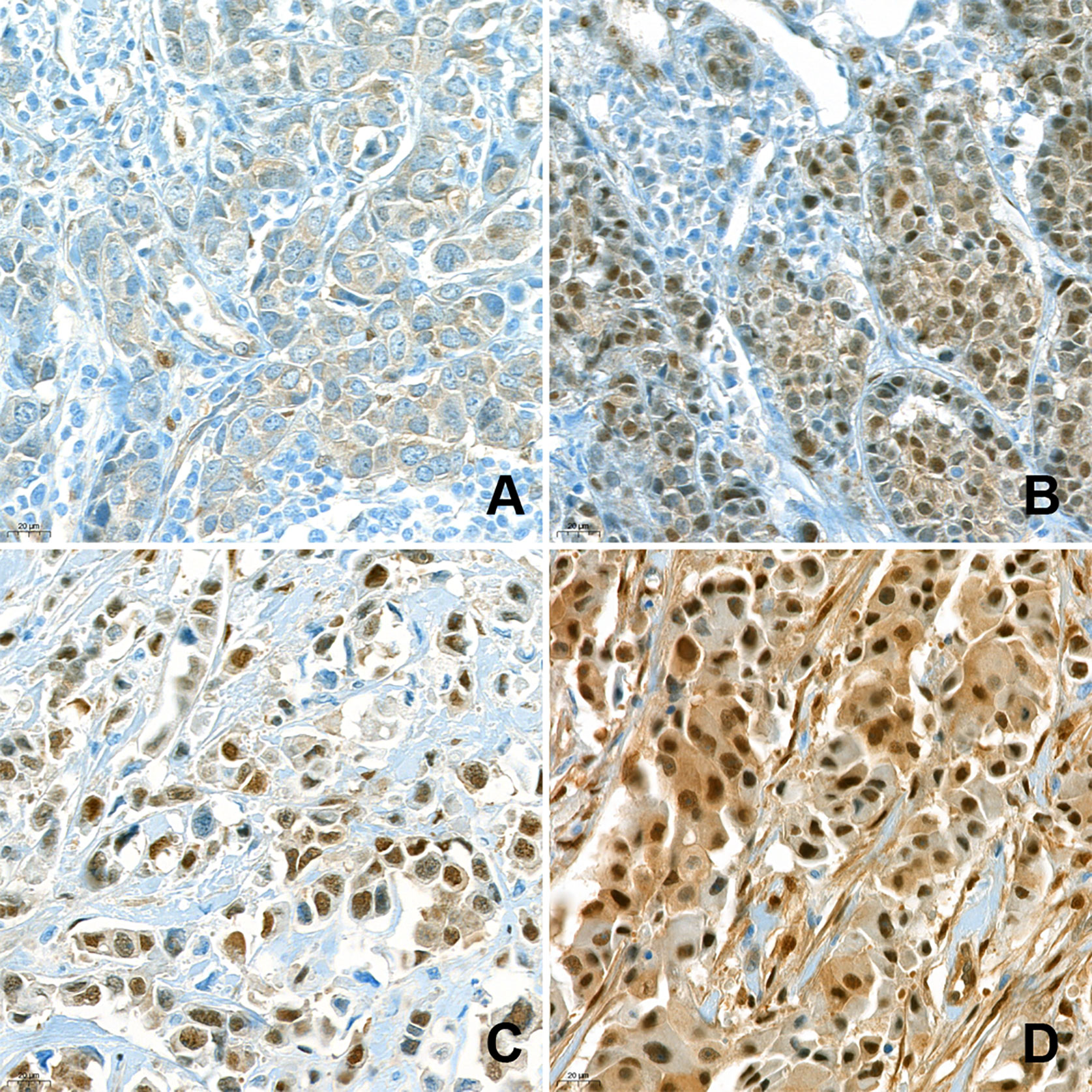
An association of a simultaneous nuclear and cytoplasmic localization of  Fra-1 with breast malignancy | BMC Cancer | Full Text
An association of a simultaneous nuclear and cytoplasmic localization of Fra-1 with breast malignancy | BMC Cancer | Full Text

IHC 2 APP: Ki-67 Nuclear Staining Analysis

Representative images of IHC staining of A) Negative staining, B)... |  Download Scientific Diagram
Representative images of IHC staining of A) Negative staining, B)... | Download Scientific Diagram

TMA immunohistochemistry showing (A) intense nuclear staining (arrow)... |  Download Scientific Diagram
TMA immunohistochemistry showing (A) intense nuclear staining (arrow)... | Download Scientific Diagram

SHON expression predicts response and relapse risk of breast cancer  patients after anthracycline-based combination chemotherapy or tamoxifen  treatment | British Journal of Cancer
SHON expression predicts response and relapse risk of breast cancer patients after anthracycline-based combination chemotherapy or tamoxifen treatment | British Journal of Cancer

Immunohistochemistry (IHC): the complete guide | Abcam

Immunohistochemistry: a): Diffuse strong nuclear staining of tumour cells  for TdT (IHC 40X); b): Diffuse strong membranous and cytoplasmic staining  for CD3 in tumour cells (IHC 40X); c): Tumour cells exhibiting diffuse
Immunohistochemistry: a): Diffuse strong nuclear staining of tumour cells for TdT (IHC 40X); b): Diffuse strong membranous and cytoplasmic staining for CD3 in tumour cells (IHC 40X); c): Tumour cells exhibiting diffuse

Immunohistochemical findings. a TFE-3 showing nuclear staining. b... |  Download Scientific Diagram
Immunohistochemical findings. a TFE-3 showing nuclear staining. b... | Download Scientific Diagram

Positive staining patterns in immunohistochemistry. (a) Membranous... |  Download Scientific Diagram
Positive staining patterns in immunohistochemistry. (a) Membranous... | Download Scientific Diagram

A. The tumor cells show strong nuclear staining (3/3) with PHLDA3... |  Download Scientific Diagram
A. The tumor cells show strong nuclear staining (3/3) with PHLDA3... | Download Scientific Diagram

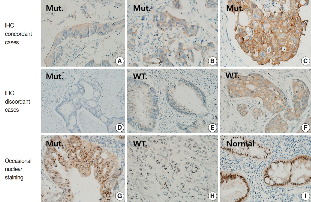
File:Main staining patterns on immunohistochemistry.jpg - Wikipedia

Journal of Pathology and Translational Medicine

A, IHC stains demonstrate diffuse positive nuclear (left) and focal... |  Download Scientific Diagram
A, IHC stains demonstrate diffuse positive nuclear (left) and focal... | Download Scientific Diagram

Histological methods for CNS

Immunohistochemical staining of WRAP53β. (A) Nuclear staining. (b)... |  Download Scientific Diagram
Immunohistochemical staining of WRAP53β. (A) Nuclear staining. (b)... | Download Scientific Diagram

Bio SB on X: "Great nuclear staining on this #IHC of our BAP1, MMab, on  mesothelioma. One of our most popular antibodies! #Antibody details here -  https://t.co/j2wulYMBCH #IHC #immunohistochemistry #antibodies #pathology  #pathart
Bio SB on X: "Great nuclear staining on this #IHC of our BAP1, MMab, on mesothelioma. One of our most popular antibodies! #Antibody details here - https://t.co/j2wulYMBCH #IHC #immunohistochemistry #antibodies #pathology #pathart
Tissue-Tek Genie | Rapid true random access IHC staining

IHC Counterstains for Enzyme-Chromogen and Fluorescent Immunostaining -  Enzo Life Sciences
IHC Counterstains for Enzyme-Chromogen and Fluorescent Immunostaining - Enzo Life Sciences

Frontiers | High Nuclear Expression of Yes-Associated Protein 1 Correlates  With Metastasis in Patients With Breast Cancer
Frontiers | High Nuclear Expression of Yes-Associated Protein 1 Correlates With Metastasis in Patients With Breast Cancer

Applied Sciences | Free Full-Text | Immunohistochemical Expression of  Wilms' Tumor 1 Protein in Human Tissues: From Ontogenesis to Neoplastic  Tissues
Applied Sciences | Free Full-Text | Immunohistochemical Expression of Wilms' Tumor 1 Protein in Human Tissues: From Ontogenesis to Neoplastic Tissues

Tissue-Tek Genie | Rapid true random access IHC staining

High magnification immunohistochemistry showing nuclear versus... |  Download Scientific Diagram
High magnification immunohistochemistry showing nuclear versus... | Download Scientific Diagram

Photograph of IHC panel: (A) IHC of TTF1 shows the nuclear positivity... |  Download Scientific Diagram
Photograph of IHC panel: (A) IHC of TTF1 shows the nuclear positivity... | Download Scientific Diagram
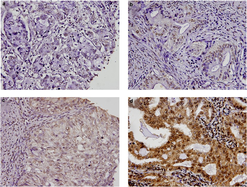
Scoring of p16INK4a immunohistochemistry based on independent nuclear  staining alone can sufficiently distinguish between endocervical and  endometrial adenocarcinomas in a tissue microarray study | Modern Pathology
Scoring of p16INK4a immunohistochemistry based on independent nuclear staining alone can sufficiently distinguish between endocervical and endometrial adenocarcinomas in a tissue microarray study | Modern Pathology