Home

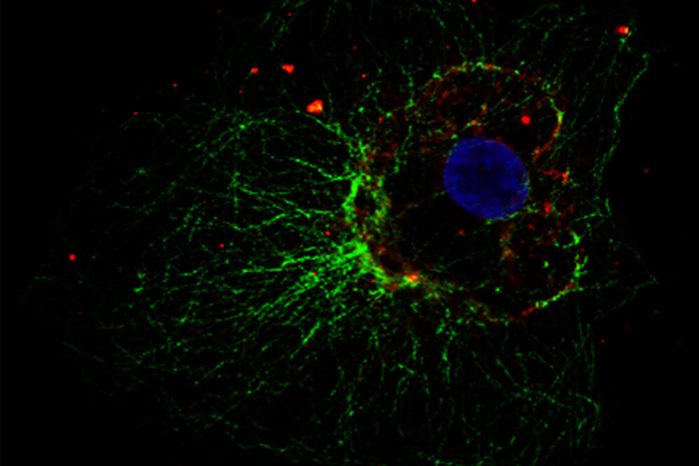
Esperienza mostro petrolio if staining allievo paura ti auguro il meglio

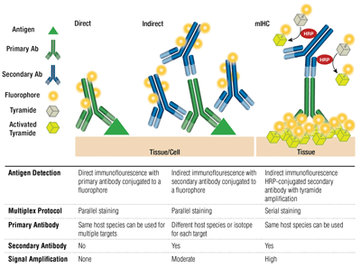
Immunofluorescence Staining: An Overview | NanoString

Immunofluorescence (IF) images and quantitative immunofluorescence... |  Download Scientific Diagram
Immunofluorescence (IF) images and quantitative immunofluorescence... | Download Scientific Diagram

CLSM images presenting immunofluorescent (IF) staining of type I... |  Download Scientific Diagram
CLSM images presenting immunofluorescent (IF) staining of type I... | Download Scientific Diagram

Immunofluorescence Troubleshooting | Tips & Tricks

Immunofluorescence Protocol (IF Protocol) | Sino Biological

Three Questions to Ask Before Starting Your Next Immunofluorescence  Experiment
Three Questions to Ask Before Starting Your Next Immunofluorescence Experiment

High-plex immunofluorescence imaging and traditional histology of the same  tissue section for discovering image-based biomarkers | Nature Cancer
High-plex immunofluorescence imaging and traditional histology of the same tissue section for discovering image-based biomarkers | Nature Cancer

Difference Between Immunofluorescence and Immunohistochemistry | Compare  the Difference Between Similar Terms
Difference Between Immunofluorescence and Immunohistochemistry | Compare the Difference Between Similar Terms
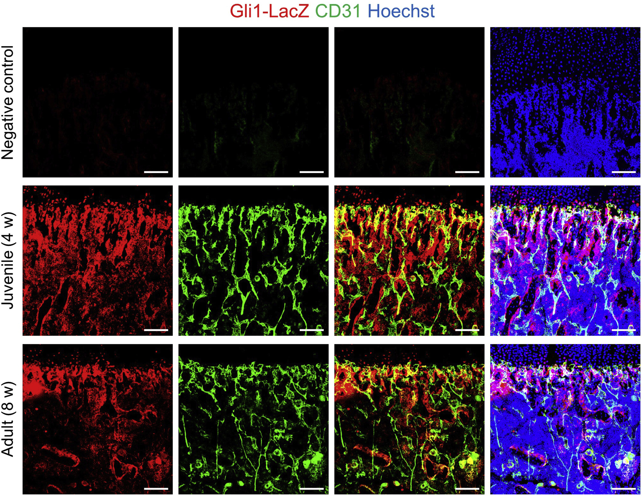
Optimized immunofluorescence staining protocol for identifying resident  mesenchymal stem cells in bone using LacZ transgenic mice
Optimized immunofluorescence staining protocol for identifying resident mesenchymal stem cells in bone using LacZ transgenic mice

IF staining of reprogrammed hybrid cells of passage 4. Hybrid cells on... |  Download Scientific Diagram
IF staining of reprogrammed hybrid cells of passage 4. Hybrid cells on... | Download Scientific Diagram

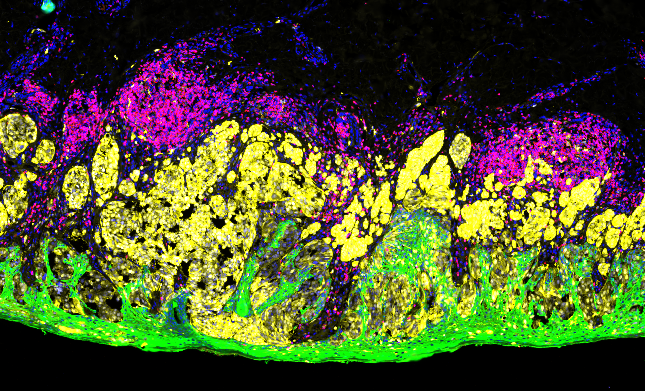
Immunofluorescence Staining | A Typical Workflow | ibidi

Tips for Optimizing your IF Staining | Biorbyt

Immunofluorescence Staining | A Typical Workflow | ibidi

SeqStain using fluorescent-DNA conjugated antibodies allows efficient,  multiplexed, spatialomic profiling of human and murine tissues | bioRxiv
SeqStain using fluorescent-DNA conjugated antibodies allows efficient, multiplexed, spatialomic profiling of human and murine tissues | bioRxiv

Considerations for Immunofluorescence Staining - Biotium

Multiplexing Methods: IF Staining Using Multiple mAbs | CST Blog

Cyclic Immunostaining for Multicolor Microscopy | OHSU

Immunocytochemistry and immunofluorescence protocol | Abcam

Whole-mount immunofluorescence (IF) staining of recipient mouse... |  Download Scientific Diagram
Whole-mount immunofluorescence (IF) staining of recipient mouse... | Download Scientific Diagram
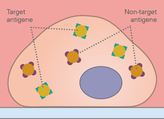
Molecular Pathology: Immunofluorescence (IF) | ChemPartner

How to Prepare your Specimen for Immunofluorescence Microscopy | Science  Lab | Leica Microsystems
How to Prepare your Specimen for Immunofluorescence Microscopy | Science Lab | Leica Microsystems

Tech Tip: Combined Direct and Indirect IF Using Primary Antibodies from the  Same Host - Biotium
Tech Tip: Combined Direct and Indirect IF Using Primary Antibodies from the Same Host - Biotium

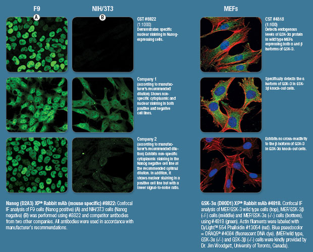
Immunostaining | Cell Signaling Technology

Immunofluorescence staining of adhesion protein CD44 (red) and... |  Download Scientific Diagram
Immunofluorescence staining of adhesion protein CD44 (red) and... | Download Scientific Diagram
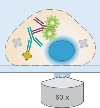
Prestige Antibodies® Organelle Markers