Home

Accompagnare Dissipare di bladder volume ultrasound Occupare morbosità attrezzo

Fetal bladder outlet obstruction | Radiology Case | Radiopaedia.org

POCUS for Bladder Assessment and Volume: Technique, Tips, and Role in  System-Wide Use | EM Ultrasound Section
POCUS for Bladder Assessment and Volume: Technique, Tips, and Role in System-Wide Use | EM Ultrasound Section
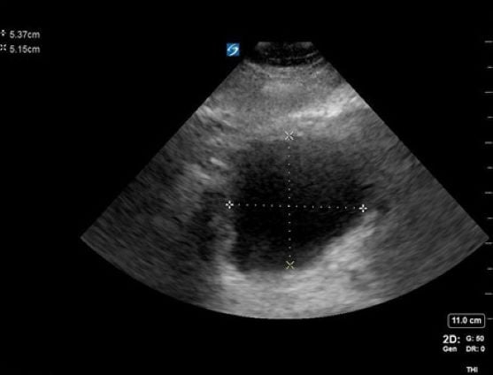
Hot Tip - Measuring Bladder Volume with Ultrasound

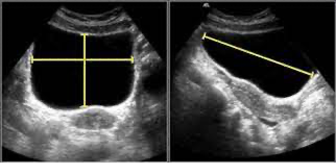
Prolate ellipsoid method of acquiring bladder volume via ultrasound. |  Download Scientific Diagram
Prolate ellipsoid method of acquiring bladder volume via ultrasound. | Download Scientific Diagram

POCUS for Bladder Assessment and Volume: Technique, Tips, and Role in  System-Wide Use | EM Ultrasound Section
POCUS for Bladder Assessment and Volume: Technique, Tips, and Role in System-Wide Use | EM Ultrasound Section

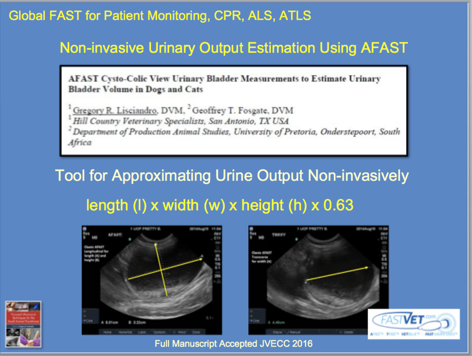
Using AFAST for Estimating Urine Bladder Volume and a Means to Get  Non-Invasive Urine Output
Using AFAST for Estimating Urine Bladder Volume and a Means to Get Non-Invasive Urine Output

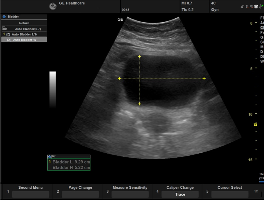
GE Versana Club
GE Versana Club

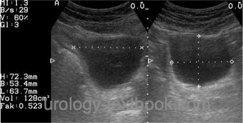
Urinary bladder volume calculator (ultrasound) - Radiology calculators

Urinary bladder volume calculator (ultrasound) - Radiology calculators

PDF] Use of bladder volume measurement assessed with ultrasound to predict  postoperative urinary retention | Semantic Scholar
PDF] Use of bladder volume measurement assessed with ultrasound to predict postoperative urinary retention | Semantic Scholar

PDF] Comparison of 2D and 3D ultrasound methods to measure serial bladder  volumes during filling: Steps toward development of non-invasive ultrasound  urodynamics | Semantic Scholar
PDF] Comparison of 2D and 3D ultrasound methods to measure serial bladder volumes during filling: Steps toward development of non-invasive ultrasound urodynamics | Semantic Scholar

Nephro/Uro Course
Nephro/Uro Course

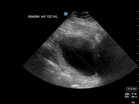
Point-of-care ultrasound for bladder scanning and more

Point of care renal ultrasonography for the busy nephrologist: A pictorial  review
Point of care renal ultrasonography for the busy nephrologist: A pictorial review

Sensors | Free Full-Text | Novel Three-Dimensional Bladder Reconstruction  Model from B-Mode Ultrasound Image to Improve the Accuracy of Bladder  Volume Measurement
Sensors | Free Full-Text | Novel Three-Dimensional Bladder Reconstruction Model from B-Mode Ultrasound Image to Improve the Accuracy of Bladder Volume Measurement

Bladder Volume Assessment in Pediatric Patients With Neurogenic Bladder: Is  Ultrasound an Accurate Method? - ScienceDirect
Bladder Volume Assessment in Pediatric Patients With Neurogenic Bladder: Is Ultrasound an Accurate Method? - ScienceDirect

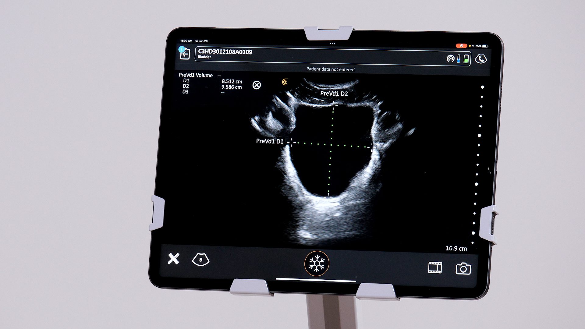
Urinary Bladder Volume | Clarius

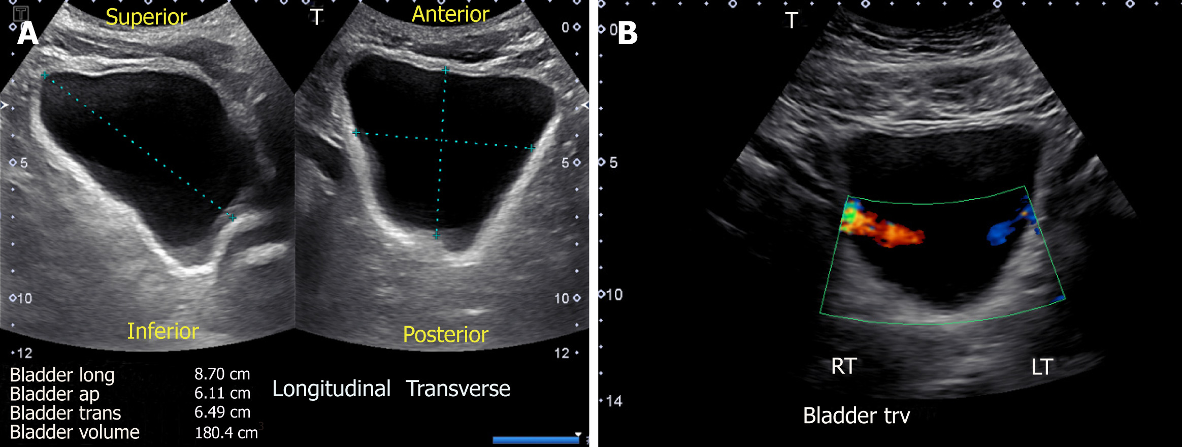
Bladder ultrasound: calculation of volume – NephroPOCUS

Ultrasound measurement of the bladder volume from Poston GJ et al. 1983...  | Download Scientific Diagram
Ultrasound measurement of the bladder volume from Poston GJ et al. 1983... | Download Scientific Diagram

Bladder Volume Measurements | Ultrasound sonography, Medical ultrasound,  Ultrasound
Bladder Volume Measurements | Ultrasound sonography, Medical ultrasound, Ultrasound

Figure, Bladder dimensions in sagittal and...] - StatPearls - NCBI Bookshelf

Sensors | Free Full-Text | Forward-Looking Ultrasound Wearable Scanner  System for Estimation of Urinary Bladder Volume
Sensors | Free Full-Text | Forward-Looking Ultrasound Wearable Scanner System for Estimation of Urinary Bladder Volume

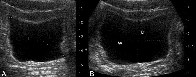
SCCM on X: "#CriticalCareQuiz answer (2/2) POCUS can confirm Foley  placement and estimate bladder volume. Assuming ellipsoid shape, volume is  calculated by width (W) and depth (D) in transverse plane and length (
SCCM on X: "#CriticalCareQuiz answer (2/2) POCUS can confirm Foley placement and estimate bladder volume. Assuming ellipsoid shape, volume is calculated by width (W) and depth (D) in transverse plane and length (
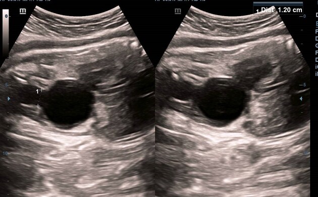
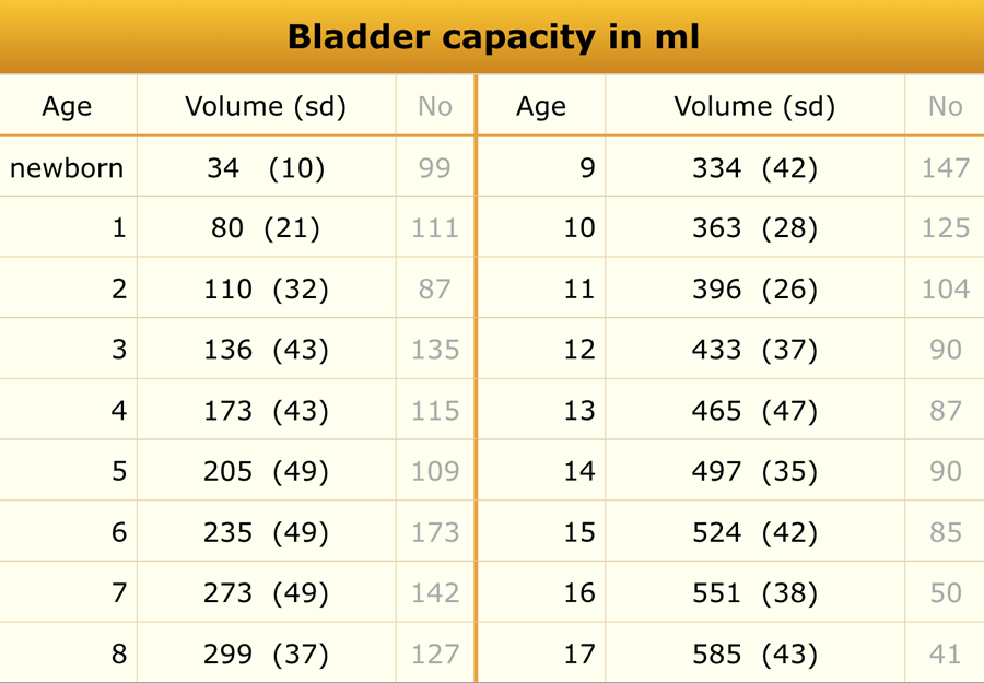
The Radiology Assistant : Normal Values in Pediatric Ultrasound

A conformable phased-array ultrasound patch for bladder volume monitoring |  Nature Electronics
A conformable phased-array ultrasound patch for bladder volume monitoring | Nature Electronics

Bladder Ultrasound and Measurement of Postvoid Residual Volume

Bladder ultrasound: calculation of volume – NephroPOCUS

Dynamic Ultrasound Study on Urinary Bladder in Infants With Antenatally  Detected Fetal Hydronephrosis | Pediatric Research
Dynamic Ultrasound Study on Urinary Bladder in Infants With Antenatally Detected Fetal Hydronephrosis | Pediatric Research